X-ray
An X-ray is a quick, painless test that uses radiation to produce images of the bones and organs of the body.

During an X-ray, a focused beam of radiation is passed through a specific area of the body, and a black-and-white image is recorded on special film or digital media.
Because the body's tissues vary in density or thickness, each tissue allows a different amount of radiation to pass through and expose the X-ray-sensitive film.
- Bones, for example, are very dense, and most of the radiation is prevented from passing through to the film. As a result, bones appear white on an X-ray
- Tissues that are less dense – such as the lungs which are filled with air – allow more of the X-rays to pass through to the film and appear on the image in shades of gray.
- The soft tissues in the body (like blood, skin, fat, and muscle) allow most of the X-ray to pass through and appear dark gray on the film or digital screen
Why do I need this test?
X-rays and other tests are part of a thorough and complete investigation of symptoms and treatment options.
The images created by X-rays of the brain, back, neck and spine help diagnose and determine treatment plans for various issues, such as fractures (breaks), tumors (abnormal masses of cells), strokes, arthritis, disc problems, deformities in the curves of the spine, osteoporosis (thinning of the bones) and infection.
X-rays are just one of the tools used by Semmes Murphey practitioners to diagnose spine, back, or neck problems. Your doctor may request other tests as well. Please see these procedure sections for additional information.
Are there any risks?
In general, X-rays are very safe and unlikely to produce any side effects. X-ray technology has been in use since its discovery and development in 1895. Today this technology is both safe and effective.
Patients will be exposed to a very small amount of radiation. The risks are minimal, but the benefits from these tests far outweigh the risks.
If a patient has a large number of X-ray exams and/or treatments over a long period of time his or her level of radiation exposure can be higher. Please alert your health care provider if you have a history of radiation exposure, or if you have had a recent barium X-ray procedure as this may interfere with obtaining an optimal exposure.
Please note that young children and developing fetuses are more sensitive to X-rays and are at higher risk for tissue damage. Radiation exposure during pregnancy may lead to birth defects. Please alert your physician if you are or think you may be pregnant. Many physicians will request a negative pregnancy test before administering an X-ray exam.
If it is necessary for you during pregnancy or your child to have an X-ray, special precautions will be made to minimize the radiation exposure to them. The health care professionals at Semmes Murphey have world renown experience treating pediatric conditions through their work with St. Jude Children’s Research Hospital and LeBonheur Children’s Hospital.
If contrast dye is used, there is a risk you may have an allergic reaction to the dye. Tell your healthcare provider if you have ever had a reaction to any contrast dye, or if you’ve had any kidney problems or are diabetic.
There may be other risks depending on your specific situation. Make sure your healthcare provider knows about all your medical issues, history and current medications before the procedure.
What should I do to prepare?
In most cases, there is very little you need to do to prepare for an X-ray.
It is important to tell the technologist if you are or may be pregnant. X-rays generally are not used on pregnant women because of the possible risk of radiation exposure to the developing baby. If it is necessary for you to have an X-ray, special precautions will be made to minimize the radiation exposure to the fetus.
Also, please notify the technologist if you are a diabetic, have an insulin pump, or if you have had a recent barium X-ray procedure as this may interfere with obtaining an optimal exposure.
Before the test begins, you may be asked to remove your clothing and put on a hospital gown.
You also will be asked to remove all jewelry and any other objects containing metal (such as eyeglasses and hair pins). This is done because metal can block the image and interfere with the test results.
What should I expect during the test?
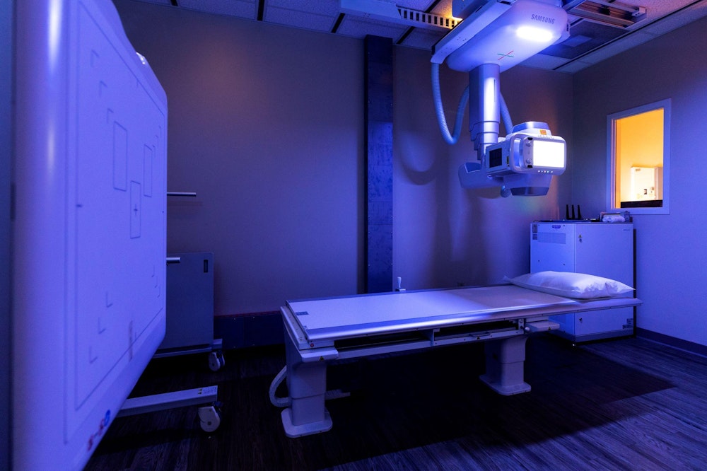
A radiology technologist, specifically trained and dedicated to administering X-ray procedures, will perform the test and guide you through the examination.
The X-ray room may be cool because air conditioning is used to keep the equipment at a constant temperature.
The test itself is painless and relatively quick. You will not feel the radiation passing through your body. It will take about 5 to 15 minutes to perform this test.
However, the manipulation of the body part being examined may cause some discomfort or pain. This is particularly true in the case of a recent injury or surgery. The radiologic technologist will use all possible comfort measures and complete the procedure as quickly as possible to reduce any discomfort or pain.
For some types of X-ray tests, a contrast medium — such as iodine or barium — is introduced into your body to provide greater detail and help highlight a specific area of your body on the X-ray image. These types of X-rays may take longer to administer.
The technologist will position your body against the X-ray film or digital screen, in a way that produces the clearest image, and focus the X-ray beam on the area to be photographed. Body parts not being imaged may be covered with a lead apron (shield) to avoid exposure to the X-rays.
Depending on the test being performed, you may be asked to stand or lie down. Most likely, the technologist will take images from different positions and vantage points.
You may be asked to change positions, move your arms or even open your mouth for an X-ray of your neck. This position moves your teeth out of the way and enables the technologist to get a clearer image of the upper bones of your neck.
He or she will ask you to be very still and hold your breath for only a few seconds while the X-rays are passed through your body. Any movement—even that which occurs when you breathe in and out—can blur the X-ray image.
The radiologic technologist will step behind a protective window while the image is taken.
After the X-rays are taken, you may be asked to wait a few minutes while the technologist makes sure the X-rays are acceptable and not blurred. If necessary, you may be asked to repeat the test to obtain a clearer image.
If your child is having an X-ray, restraints or other techniques may be used to keep a them still. These won't harm your child. They will prevent your child from moving and the need for a repeat procedure.
The physicians at Semmes Murphey Clinic have a world renown reputation in pediatric surgery and treatment based on their many years’ experience treating patients at both LeBonheur Children’s Hospital and St. Jude Children’s Research Hospital. Your doctor will help you prepare your child for this test and any phase of their treatment.
You may be allowed to stay with your child during the test. If you do, you'll likely be asked to wear a lead apron to shield you from unnecessary exposure.
After an X-ray, you generally can resume normal activities. Routine X-rays usually have no side effects. However, if you receive contrast medium before the test, drink plenty of fluids afterwards to help rid your body of it.
If you have a contrast medium, ask your doctor about any signs and symptoms to watch for.
When will I receive my results?
X-rays are saved digitally which can be viewed on-screen within minutes. A radiologist (a doctor who specializes in evaluating X-rays and other radiology procedures) will interpret the X-rays and report the test results to your doctor.
If your appointment is the same day as your test, your physician may review them with you at that time. Otherwise, most test results are reviewed with you at your next scheduled appointment.
This information was provided by the specialists at Semmes Murphey Clinic. Readers are encouraged to research trustworthy organizations for information. Please talk with your physician for websites and sources that will enhance your knowledge and understanding of this issue and its treatments.

Experience the Semmes Murphey Difference for Yourself
Request an Appointment